41 cell drawing with labels
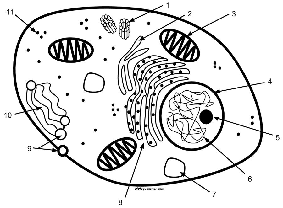
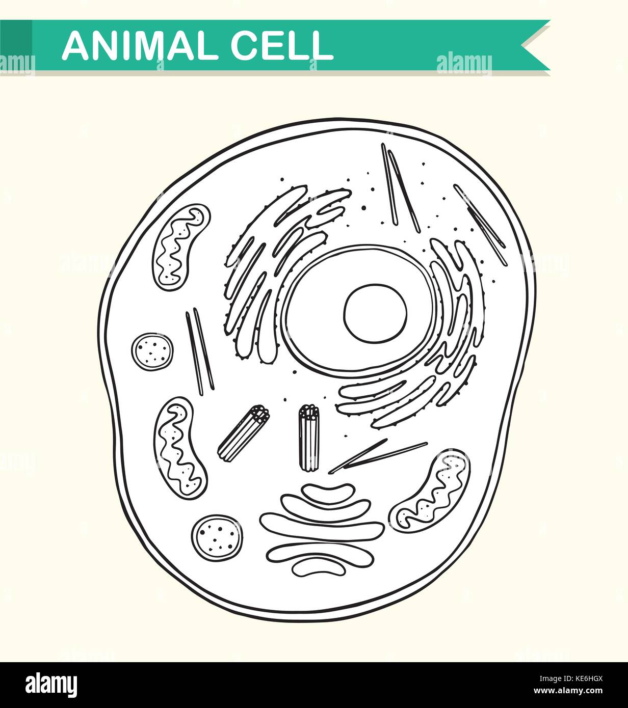
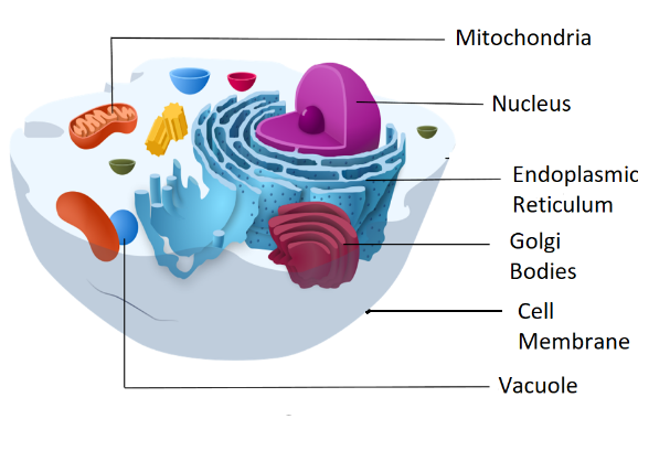
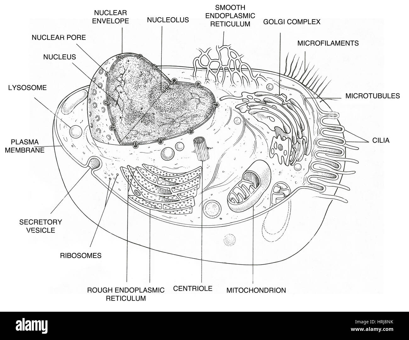
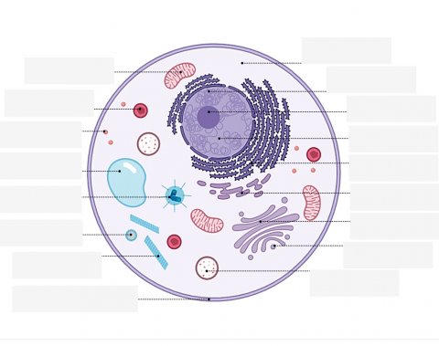
Lab Drawings - BIOLOGY FOR LIFE Positioning: Center drawing on the page. Do not draw in a corner. This will leave plenty of room for the addition of labels. Size: Make a large, clear drawing; it should occupy at least half a page. Labels: Use a ruler to draw straight, horizontal lines. The labels should form a vertical list. All labels should be printed (not cursive). A Labeled Diagram of the Animal Cell and its Organelles As observed in the labeled animal cell diagram, the cell membrane forms the confining factor of the cell, that is it envelopes the cell constituents together and gives the cell its shape, form, and existence. Cell membrane is made up of lipids and proteins and forms a barrier between the extracellular liquid bathing all cells on the exterior ...
draw and label plant cell - TeachersPayTeachers This worksheet includes both an extensive animal and plant cell diagram to label. The animal cell includes 17 organelles, and the plant cell includes 20 organelles for students to label and color. There is also a 4 page graphic organizer (chart) that includes a drawing of each of the organelles in alphabetical order.

Cell drawing with labels
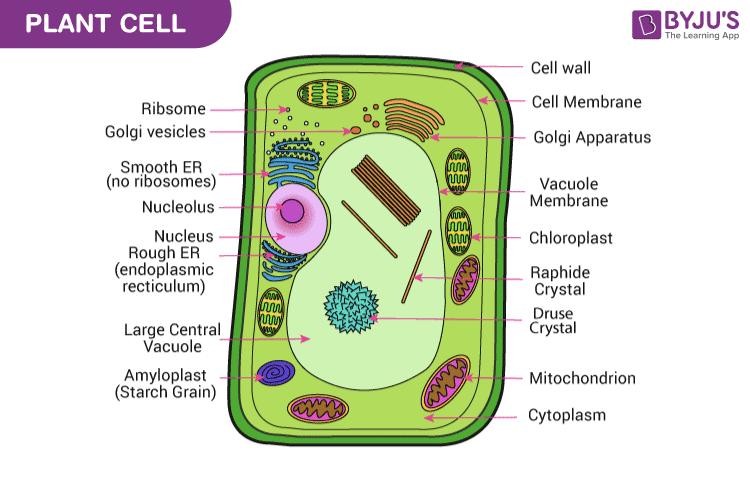
Labeled Plant Cell With Diagrams | Science Trends The parts of a plant cell include the cell wall, the cell membrane, the cytoskeleton or cytoplasm, the nucleus, the Golgi body, the mitochondria, the peroxisome's, the vacuoles, ribosomes, and the endoplasmic reticulum. Parts Of A Plant Cell The Cell Wall Let's start from the outside and work our way inwards. Label animal cell - Teaching resources - Wordwall 10000+ results for 'label animal cell'. Label Animal Cell Organelles Labelled diagram. by Britter. Label Animal Cell Organelles Labelled diagram. by Mbauer. Label Plant and Animal Cell Labelled diagram. by Catherine34. Animal Cell Label Labelled diagram. by Taraabbott. Creating File Folder Labels In Microsoft Word - Worldlabel.com 2. Pick a shape, and then you’ll get a plus-sign-like drawing cursor. Draw the shape to fill the label cell. 3. If your shape doesn’t perfectly land within the area you want it, click on the little handles in the frame surrounding the shape to resize it to fit. 4.
Cell drawing with labels. Cell Junctions - Molecular Biology of the Cell - NCBI Bookshelf Specialized cell junctions occur at points of cell-cell and cell-matrix contact in all tissues, and they are particularly plentiful in epithelia. Cell junctions are best visualized using either conventional or freeze-fracture electron microscopy (discussed in Chapter 9), which reveals that the interacting plasma membranes (and often the underlying cytoplasm and the intervening … Free Cell Diagram Software with Free Templates - EdrawMax - Edrawsoft An animal cell diagram describes a cell structure enclosed by a plasma member, and it has a nucleus with a membrane and organelles. Neuron Diagram A neuron diagram describes the three parts of a Neuron: dendrites, an axon, a cell body, or soma. Cell Membrane Diagram What's new in think-cell :: think-cell For the table cells for status, choose Checkbox in the cell content control and activate Use Excel Cell Border.The status will now be shown as checkboxes, with content determined by the Excel cell. In the Excel cell, use v, o or 1 for “check”; x or 2 for “cross”; Space or 0 for an unchecked box. Moreover, the border lines of all cells in the table are now controlled by those set in Excel. CELL MEMBRANE LABEL Diagram | Quizlet identifies or labels the cell. Receptor protein. receives information. Heads. part of the phospholipid that loves water (hydrophili) - points to the most outside and inside of cell. Tails. part of phospholipid that hates water (hydrophobic); points to the interior or Inside. Phospholipid Bilayer. 2 layers of fat - tails point in toward each ...
Cell Cycle Diagram Labeled | EdrawMax Cell Cycle Diagram Labeled Use Template 1 0 Report Publish time:03-02-2022 Tag: science diagram The following labeled diagram illustrates the cell cycle. As you can see in the labeled diagram here, the cell cycle is an ordered series of events involving cell growth and cell division that produces two new daughter cells. Cell Size and Scale - University of Utah Smaller cells are easily visible under a light microscope. It's even possible to make out structures within the cell, such as the nucleus, mitochondria and chloroplasts. Light microscopes use a system of lenses to magnify an image. The power of a light microscope is limited by the wavelength of visible light, which is about 500 nm. Labels - Etsy Cell Phone Accessories ... Drawing & Drafting Photography Collage ... 250 cotton labels, cotton tags, fabric label, printed labels, printing tag, natural labels, end fold labels, cotton tags 5 out of 5 stars (1,553) Star Seller $ 65.00. ad vertisement by Etsy ... Plant Cell Diagram drawing CBSE || easy way || Labeled ... - YouTube these are some parts of the plant cell: list of plant cell organelles cell wall cytoskeleton cell (plasma) membrane plasmodesmata the cytoplasm plastids plant vacuoles mitochondria endoplasmic...
How to Draw an Animal Cell: 11 Steps (with Pictures) - wikiHow Draw a simple circle or oval for the cell membrane. The cell membrane of an animal cell is not a perfect circle. You can make the circle misshapen or oblong. The important part is that it does not have any sharp edges. [1] Also know that the membrane is not a rigid cell wall like in plant cells. Structure of Cell: Definition, Types, Diagram, Functions - Embibe Structure of Cell: Cell is the basic functional unit that makes up all living organisms.All organisms, including ourselves, start life as a single cell called the egg. Cells are small microscopic units that perform all essential functions of life and are capable of independent existence. How to draw an animal cell - labeled science diagram - YouTube Download a free printable outline of this video and draw along with us: you for watching. Please ... Red Blood Cell Diagram Labeled stock illustrations Red Blood Cell Diagram Labeled stock illustrations View red blood cell diagram labeled videos Browse 19 red blood cell diagram labeled stock illustrations and vector graphics available royalty-free, or start a new search to explore more great stock images and vector art. Newest results Human gas exchange system vector illustration. Oxygen travel...
Animal Cells: Labelled Diagram, Definitions, and Structure - Research Tweet The endoplasmic reticulum (s) are organelles that create a network of membranes that transport substances around the cell. They have phospholipid bilayers. There are two types of ER: the rough ER, and the smooth ER. The rough endoplasmic reticulum is rough because it has ribosomes (which is explained below) attached to it.
White Blood Cell Diagram Labeled Pictures, Images and Stock Photos White blood cell types labeled examples educational vector illustration. Isolated WBC closeup scheme with neutrophil, eosinophil, monocyte, basophil and lymphocytes differences comparison collection. Neutrophil vector illustration. Medical educational scheme with... Neutrophil vector illustration.
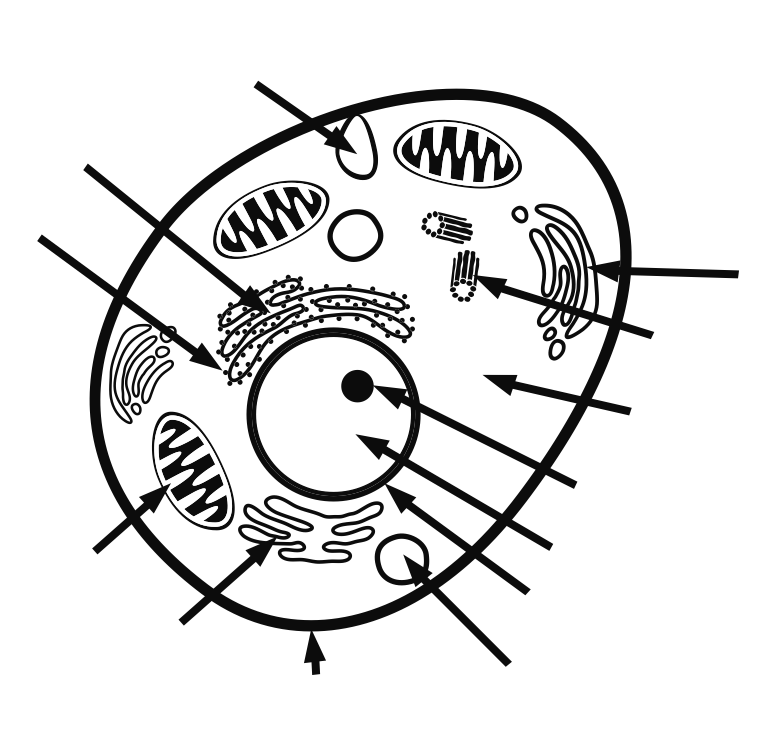
Learn the parts of a cell with diagrams and cell quizzes For this exercise we'll start with an image of a cell diagram ready labeled. Study this and make sure that you're clear about which structure is found where. Cell diagram unlabeled It's time to label the cell yourself! As you fill in the cell structure worksheet, remember the functions of each part of the cell that you learned in the video.
Cells Diagram | Science Illustration Solutions - Edrawsoft Edraw software offers you lots of symbols used in cells diagram like cell structure, paramecium, squamous cell, cell division, bacteria, cell membrane, eggs, sperm, zygote, an animal cell, SARS, tobacco mosaic, adenovirus, coliphage, herpesvirus, AIDS, pollen, plant cell model, onion tissue, etc. Cells Diagram Examples
Drawing & Labeling a Diagram of a Electrochemical Cell Labeled Electrochemical Cell 'If we draw and label our electrochemical cell and include the direction of electron flow, we should get something like this,' said Xavier.
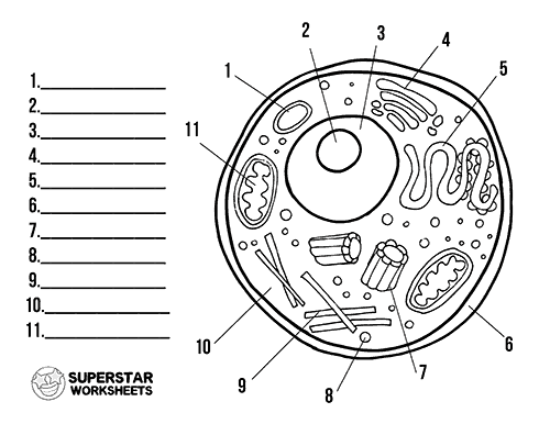
Animal Cell - Free printable to label + Color -kidCourses.com Can you label and color these important parts of the animal cell?. NUCLEUS control center for cell (cell growth, cell metabolism, cell reproduction). NUCLEOLUS synthesizes rRNA. RIBOSOMES the site of protein building, this is where translation takes place (mRNA in language of nucleic acids is translated into the language of amino acids). RER (Rough Endoplasmic Reticulum) synthesizes proteins ...
Cell: Structure and Functions (With Diagram) - Biology Discussion Eukaryotic Cells: 1. Eukaryotes are sophisticated cells with a well defined nucleus and cell organelles. 2. The cells are comparatively larger in size (10-100 μm). 3. Unicellular to multicellular in nature and evolved ~1 billion years ago. 4. The cell membrane is semipermeable and flexible. 5. These cells reproduce both asexually and sexually.
A Well-labelled Diagram Of Animal Cell With Explanation - BYJUS The animal cell diagram is widely asked in Class 10 and 12 examinations and is beneficial to understand the structure and functions of an animal. A brief explanation of the different parts of an animal cell along with a well-labelled diagram is mentioned below for reference. Also Read Different between Plant Cell and Animal Cell
Custom Color-Coded Maps – shown on Google Maps 2 days ago · 1. In Google Sheets, create a spreadsheet with 4 columns in this order: County, StateAbbrev, Data* and Color • Free version has a limit of 1,000 rows • Map data will be read from the first sheet tab in your Google Sheet • If you don't have a Google Sheet, create one by importing from Excel or a .csv file • The header of the third column will be used as the map …
Labels - Etsy Check out our labels selection for the very best in unique or custom, handmade pieces from our stickers, labels & tags shops.
PDF Human Cell Diagram, Parts, Pictures, Structure and Functions Sickle cell anemia (SCA) is an inherited anemic condition that appears due to a defect in the gene coding for hemoglobin (HbS). Owing to the mutation, RBCs become sickle-shaped (crescent- shaped). The lifespan of these defective red blood cells are also greatly reduced.
Microsoft Word - Work together on Word documents Collaborate for free with an online version of Microsoft Word. Save documents in OneDrive. Share them with others and work together at the same time.
Label Cells Teaching Resources | Teachers Pay Teachers Plant and Animal Cells Color and Label Parts by Rulers and Pan Balances 4.7 (43) $1.00 PDF The set includes two versions of a plant cell to color and label and two versions of an animal cell to color and label.
Human Cell Draw, Label, Define.docx - Human Cell Draw,... Human Cell Draw, Label, and Define Golgi Apparatus - A cell organelle found in the cytoplasm that synthesizes carbohydrates and packages materials for secretion from the cell. Chromatin - A thread like structure of genetic material from a cell that is not dividing Nucleus-Membrane part of cell that contains the hereditary material in chromosomes. ...
Animal Cell Diagram Labeled | EdrawMax Template According to the animal cell labeled diagram, some of the cell organelles of an animal cell are the cell membrane, Cytosol, Cytoskeleton, nucleus, Ribosomes, Endoplasmic Membrane, Vesicles, Mitochondria, and more. Creating such labeled diagrams will teach your young students more about animal cells.
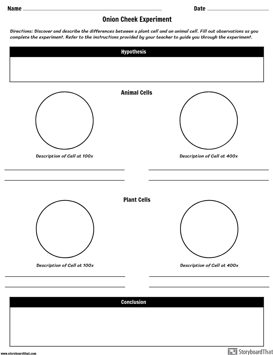
Label Cell Parts | Plant & Animal Cell Activity | StoryboardThat Create a cell diagram with each part of plant and animal cells labeled. Include descriptions of what each organelle does. Click "Start Assignment". Find diagrams of a plant and an animal cell in the Science tab. Using arrows and Textables, label each part of the cell and describe its function.
Personalized Address Labels - Etsy Check out our personalized address labels selection for the very best in unique or custom, handmade pieces from our address & shipping labels shops.
2,146 Red blood cell diagram Images, Stock Photos & Vectors | Shutterstock Find Red blood cell diagram stock images in HD and millions of other royalty-free stock photos, illustrations and vectors in the Shutterstock collection. Thousands of new, high-quality pictures added every day.
School Labels - Etsy Check out our school labels selection for the very best in unique or custom, handmade pieces from our stickers, labels & tags shops.
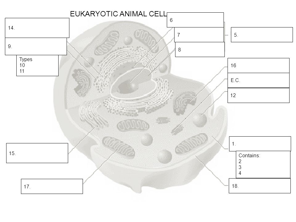
Label the Animal Cell: Level 2 | Worksheet | Education.com Students take a deeper dive into the structure of animal cells in this middle grades life science worksheet! In Label the Animal Cell: Level 2, students will use a word bank to label the parts of a cell in an animal cell diagram. For added enrichment, have students assign a color to each of the organelles and then color in the diagram.
Labeling a Cell Diagram | Quizlet Labeling a Cell + − Flashcards Learn Test Match Created by MissEStrauss Terms in this set (15) Nucleus The command center of the cell. It controls cell's activity. Cytoplasm A jelly-like substance that fills the cell. All of the cell's organelles are located here. Vacuoles The storage facilities for the cell.
Two-Level Axis Labels (Microsoft Excel) - tips 16/04/2021 · Excel automatically recognizes that you have two rows being used for the X-axis labels, and formats the chart correctly. (See Figure 1.) Since the X-axis labels appear beneath the chart data, the order of the label rows is reversed—exactly as mentioned at the first of this tip. Figure 1. Two-level axis labels are created automatically by Excel.
Creating File Folder Labels In Microsoft Word - Worldlabel.com 2. Pick a shape, and then you’ll get a plus-sign-like drawing cursor. Draw the shape to fill the label cell. 3. If your shape doesn’t perfectly land within the area you want it, click on the little handles in the frame surrounding the shape to resize it to fit. 4.
Label animal cell - Teaching resources - Wordwall 10000+ results for 'label animal cell'. Label Animal Cell Organelles Labelled diagram. by Britter. Label Animal Cell Organelles Labelled diagram. by Mbauer. Label Plant and Animal Cell Labelled diagram. by Catherine34. Animal Cell Label Labelled diagram. by Taraabbott.
Labeled Plant Cell With Diagrams | Science Trends The parts of a plant cell include the cell wall, the cell membrane, the cytoskeleton or cytoplasm, the nucleus, the Golgi body, the mitochondria, the peroxisome's, the vacuoles, ribosomes, and the endoplasmic reticulum. Parts Of A Plant Cell The Cell Wall Let's start from the outside and work our way inwards.


![Expert Verified] draw the diagrams of plant cell and animal ...](https://hi-static.z-dn.net/files/df9/6e9f34500f9d99c9b11f184891378fc0.jpg)










Post a Comment for "41 cell drawing with labels"