44 labelled femur
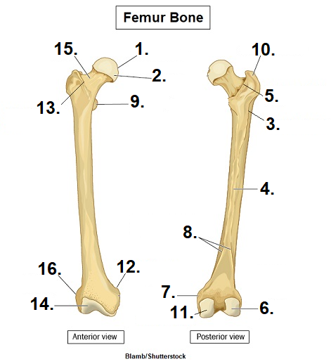
Femur Bone Anatomy: Quiz, Labeled Diagram, Skeletal System Parts - EZmed The femur is a type of long bone located in the thigh and is the largest bone of the skeletal system. There was a previous EZmed post (see below) on the anatomy of the femur where we labeled all of the main parts of the bone on a color-coded diagram. For the step-by-step video and blog post that walks through the anatomy of the femur, click below! Femur Anatomy Quiz - Registered Nurse RN Femur quiz for anatomy and physiology! This unlabeled quiz of the femur will test your knowledge on how to label the structures of this bone. You will be required to label the fovea capitis, lateral epicondyle, linea aspera, intertrochanteric line, intertrochanteric crest, adductor tubercle, greater trochanter, lesser trochanter etc.
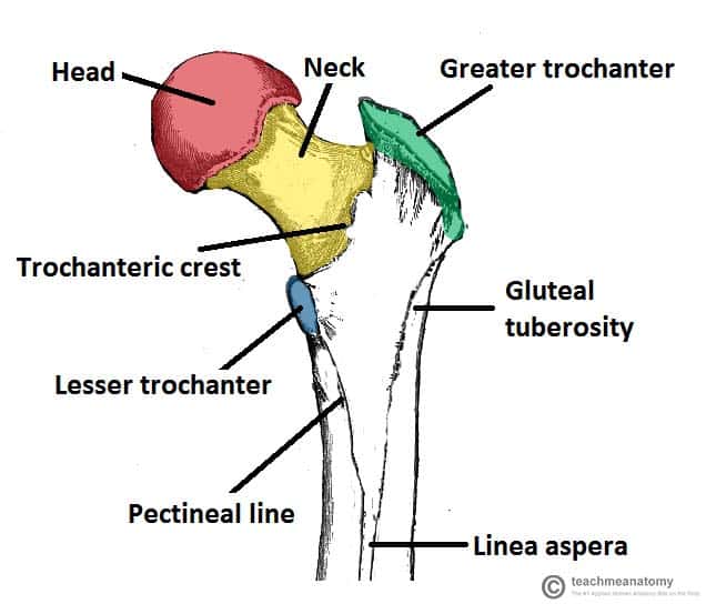
Learn femur anatomy fast with these femur quizzes | Kenhub Labeled overview of the femur bone. Femur bone unlabeled. Hopefully by this stage, all the parts of the femur have begun to slot into place in your memory. So there's only one thing left to do - put it to the test! To begin, try our femur labeling quiz by downloading the free PDF below.
Labelled femur
labeled femur Flashcards | Quizlet labeled femur. Term. 1 / 18. Articular Cartilage. Click the card to flip 👆. Definition. 1 / 18. ... Click the card to flip 👆. Labeled Femur Illustrations, Royalty-Free Vector Graphics & Clip Art ... Choose from Labeled Femur stock illustrations from iStock. Find high-quality royalty-free vector images that you won't find anywhere else. Femur Bone Anatomy: Labeled Diagram and Quiz - EZmed Femur Bone Anatomy. The femur is a type of long bone located in the thigh and is the largest bone of the skeletal system. The femur and/or hip may fracture secondary to trauma, so understanding the femur bone anatomy is important. The anatomy of the femur can be divided into proximal, central, distal, and posterior parts.
Labelled femur. Femur drawing labelled diagram, Picture #1390560 femur drawing blank Original file at image/png format. Also Femur drawing labelled diagram available at PNG transparent variant. Look at links below to get more options for getting and using clip art; Resolution 250 x 228; File Size 0.03 Kb; Uploaded 1459 days ago; Format image/png Femur (Thighbone): Anatomy, Function & Common Conditions - Cleveland Clinic Femur. The femur is the longest, strongest bone in your body. It plays an important role in how you stand, move and keep your balance. Femurs usually only break from serious traumas like car accidents. But if your bones are weakened by osteoporosis, you have an increased risk for fractures you might not even know about. Appointments 216.444.2606. Femur: Anatomy, Function, and Treatment - Verywell Health The femur is the largest and strongest bone in the human body. 1 It is commonly known as the thigh bone (femur is Latin for thigh) and reaches from the hip to the knee. The femur is extremely hard and not easy to break. A broken thigh bone is one of the few simple fractures that can be considered life-threatening because it can cause ... femur | Definition, Function, Diagram, & Facts | Britannica femur, also called thighbone, upper bone of the leg or hind leg. The head forms a ball-and-socket joint with the hip (at the acetabulum), being held in place by a ligament (ligamentum teres femoris) within the socket and by strong surrounding ligaments. In humans the neck of the femur connects the shaft and head at a 125° angle, which is efficient for walking. A prominence of the femur at the ...
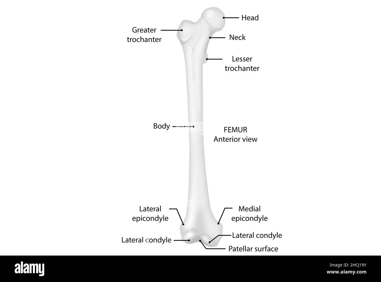
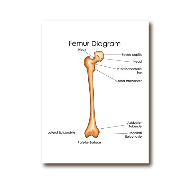
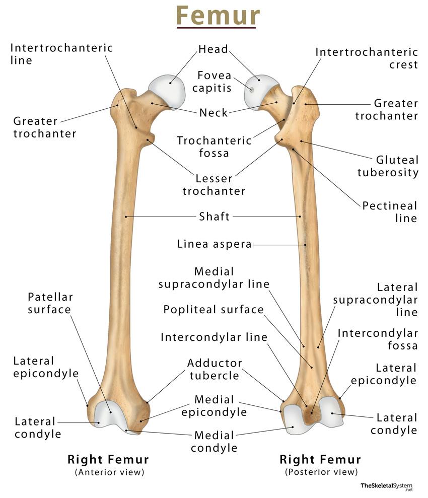
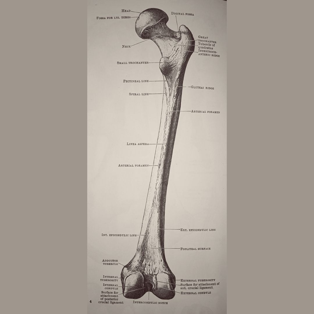
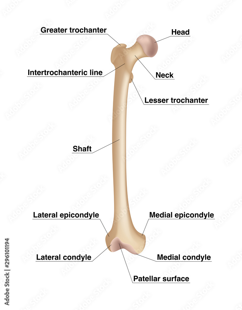
Femur drawing labelled diagram, Picture #1390558 femur drawing blank ... Original file at image/png format. Also Femur drawing labelled diagram available at PNG transparent variant. Look at links below to get more options for getting and using clip art; Resolution 512 x 711; File Size 0.15 Kb; Uploaded 1467 days ago; Format image/png Femur bone anatomy: Proximal, distal and shaft | Kenhub The femoral apophyses are prominent protrusions found on the proximal aspect of the femur. The lateral and larger of the two apophyses is the greater trochanter; its proximal edge is roughly a hand's breadth inferior to the pubic tubercle on the pubis.The great trochanter is roughly quadrangular and extends from the superior aspect of the junction of the neck and shaft of the femur. Femur Bone - Anterior and Posterior Markings | GetBodySmart The femur ( os femoris) extends from the hip to the knee and is the longest and strongest bone in the body. Forming the midportion of the femur is a long cylindrical shaft, which arches or curves anteriorly. At its proximal end, the spherical head of the femur articulates with the acetabulum ( hip socket) of the os coxa ( hip bone) to form the ... Femur Labeled Diagram| EdrawMax Template The following labeled diagram shows the Right Femur from Anterior View and Posterior View. As shown in the following labeled diagram, the femur is a type of long bone located in the thigh and the largest human anatomy bone. For better understanding, we have divided the femur into multiple parts: proximal, central, distal, and posterior parts.
Femur Bone Anatomy: Skeletal System Lower Limb [Labeled Diagram] Anatomy of the femur, or thigh bone, made easy using a colored labeled diagram and drawing. Skeletal system anatomy of lower limb for nursing, medical learne... Femur Anatomy, Diagram & Definition | Body Maps - Healthline Femur. The femur is the only bone located within the human thigh. It is both the longest and the strongest bone in the human body, extending from the hip to the knee. Important features of this ... Femur - Wikipedia The femur (/ ˈ f iː m ər /; pl. femurs or femora / ˈ f ɛ m ər ə /), or thigh bone, is the proximal bone of the hindlimb in tetrapod vertebrates.The head of the femur articulates with the acetabulum in the pelvic bone forming the hip joint, while the distal part of the femur articulates with the tibia (shinbone) and patella (kneecap), forming the knee joint.By most measures the two (left ... Femur Labeling Quiz - PurposeGames.com This is an online quiz called Femur Labeling. There is a printable worksheet available for download here so you can take the quiz with pen and paper. Your Skills & Rank. Total Points. 0. Get started! Today's Rank--0. Today 's Points. One of us! Game Points. 11. You need to get 100% to score the 11 points available.
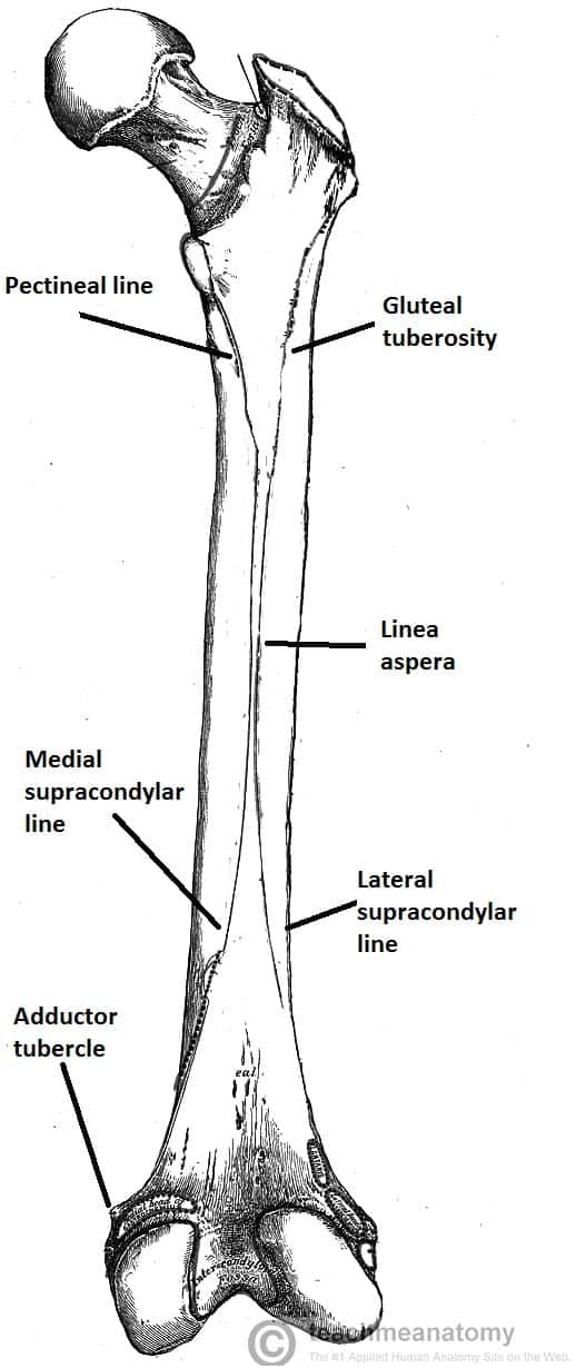
The Femur - Proximal - Distal - Shaft - TeachMeAnatomy
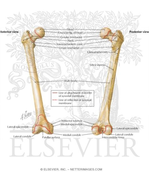
The Femur - Proximal - Distal - Shaft - TeachMeAnatomy The Femur. The femur is the only bone in the thigh and the longest bone in the body. It acts as the site of origin and attachment of many muscles and ligaments, and can be divided into three parts; proximal, shaft and distal. In this article, we shall look at the anatomy of the femur - its attachments, bony landmarks, and clinical correlations.

Consider the diagram given above. Parts labelled as 'A', 'B ...
Labeled Femur Stock Photos, Pictures & Royalty-Free Images - iStock 3D Rendering of male pelvis, hip, leg bones and ligaments labeled on a white background. Front view. Shin splints vector illustration. Leg muscle sport trauma and... Shin splints vector illustration. Leg muscle sport trauma and bone pain labeled diagram. Isolated femur, patella, fibula, tibia and foot bones with shown injury location.

Femur Bone Anatomy: Labeled Diagram, Quiz, Color-Coded Parts ...
Femur Bone Anatomy: Labeled Diagram and Quiz - EZmed Femur Bone Anatomy. The femur is a type of long bone located in the thigh and is the largest bone of the skeletal system. The femur and/or hip may fracture secondary to trauma, so understanding the femur bone anatomy is important. The anatomy of the femur can be divided into proximal, central, distal, and posterior parts.
Femur
Labeled Femur Illustrations, Royalty-Free Vector Graphics & Clip Art ... Choose from Labeled Femur stock illustrations from iStock. Find high-quality royalty-free vector images that you won't find anywhere else.
The Femur - Proximal - Distal - Shaft - TeachMeAnatomy
labeled femur Flashcards | Quizlet labeled femur. Term. 1 / 18. Articular Cartilage. Click the card to flip 👆. Definition. 1 / 18. ... Click the card to flip 👆.
Femur, anterior view, anatomy, human body Stock Photo - Alamy

Pencil-femur-labelled | Medical-Artist.com

Femur Bone Anatomy: Labeled Diagram, Quiz, Color-Coded Parts ...

Femur labeled diagram hi-res stock photography and images - Alamy

The femur | Human body anatomy, Body anatomy, Human anatomy ...

Femur Bone Anatomy: Labeled Diagram, Quiz, Color-Coded Parts ...
AkuBudakGunung™ on Twitter: "Tulang kt LUTUT ada 4 : 1) femur ...
Learn femur anatomy fast with these femur quizzes | Kenhub
Femur: Anterior View
Manusia Femur Tulang Diagram Poster Dokter Kantor Dinding ...

Femur, artwork - Stock Image - C020/9159 - Science Photo Library
Femur Anatomy Quiz

Anatomy In Motion - Femur: The femur is the only bone of the ...
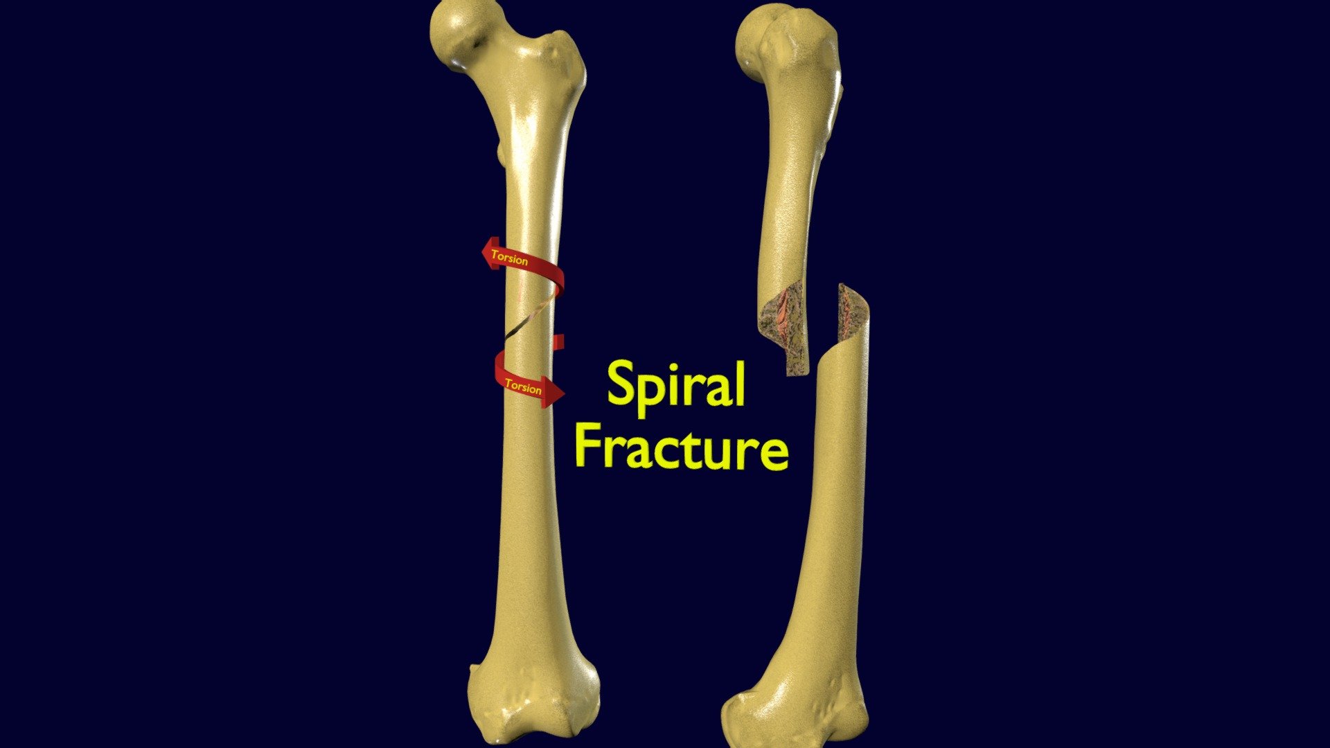
Spiral Fracture skeleton labelled femur - Buy Royalty Free 3D ...

Left: The initial label configuration for the knee joint. The ...

Femur Photograph by Asklepios Medical Atlas | Fine Art America

Femur Labeled Diagram | Quizlet

Anatomi Desain Label Sistem Kerangka Manusia Foto Stok ...

Locations of thin sections used in the auto-control study. A ...

Measurements on the large femur from Un-named Cave JF 155 ...
الفترة المحيطة بالجراحة بشكل يومي غائم مانغا السداد تدحرج ...

Anterior View - Femur Diagram | Quizlet

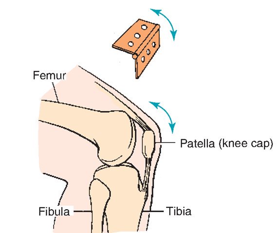
Anatomy of the knee joint, labelled, eps8. | CanStock

Femur labeled diagram hi-res stock photography and images - Alamy

Femur Bone Anatomy: Labeled Diagram, Quiz, Color-Coded Parts ...

File:Fémur insertions musculaires face postérieure.png ...
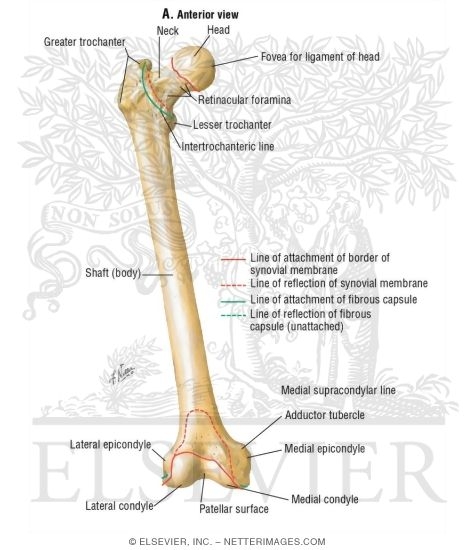
Osteology of the Femur
2,545 Labelled bones leg Images, Stock Photos & Vectors ...

File:Labelled Femur Q Angle.jpg - Wikimedia Commons

Femur labeled diagram hi-res stock photography and images ...

Femur and Humerus Diagram | Quizlet

Lecture 7 Lower Limb: Hip and Anterior and Medial Thigh ...

Consider the parts labelled as a, b, c, d and e respectively ...

Gaussian curvature sign regions on typical femurs. (a) Labels ...
The Old Operating Theatre Museum & Herb Garret en Twitter ...

Femur labeled diagram hi-res stock photography and images - Alamy

femur | Definition, Function, Diagram, & Facts | Britannica
Femur. Realistic drawing showing thigh bone with inscriptions ...

File:Human leg bones labeled.svg - Wikimedia Commons
Post a Comment for "44 labelled femur"