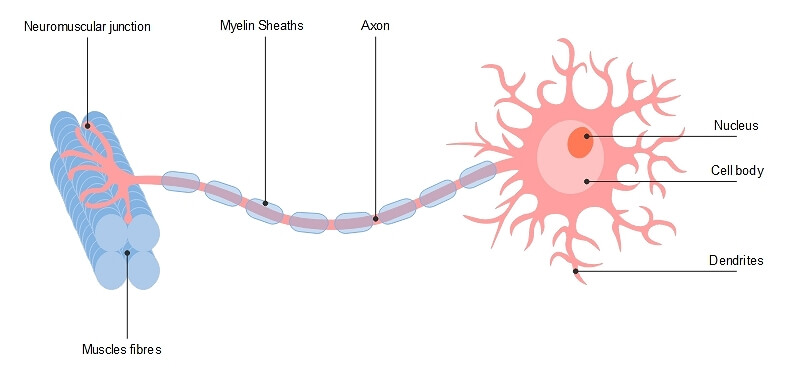
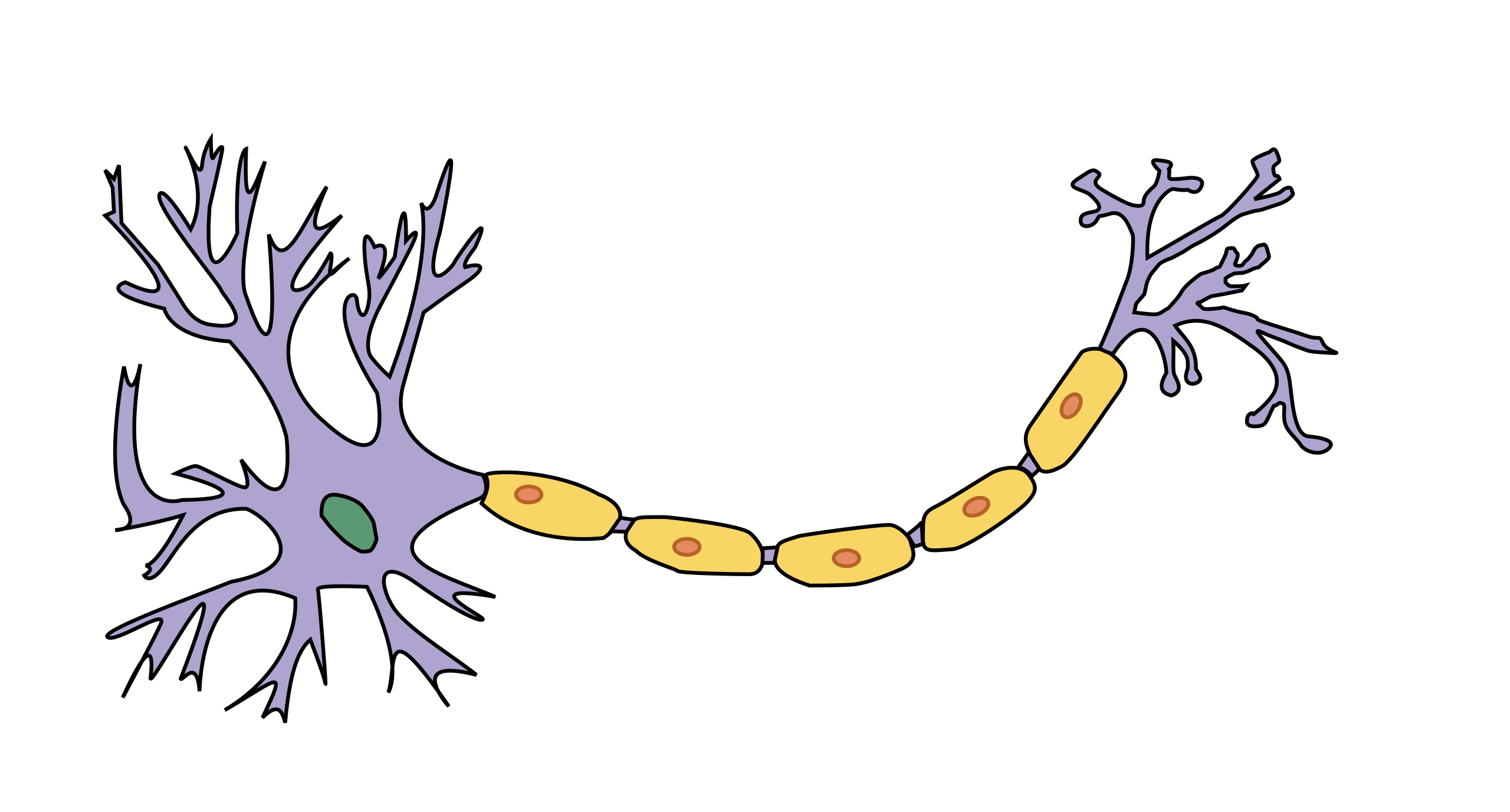
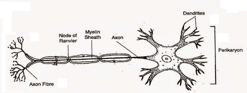
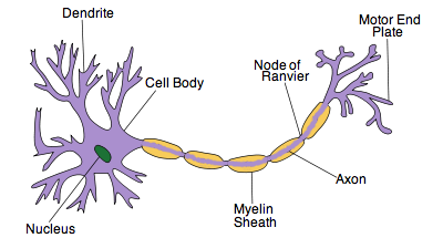
40 draw a labeled diagram of neuron
reflex arc - YouTube WebAbout Press Copyright Contact us Creators Advertise Developers Terms Privacy Policy & Safety How YouTube works Test new features Press Copyright Contact us Creators ... Andrew File System Retirement - Technology at MSU WebInformation technology resources, news, and service information at MSU. It is maintained by IT Services and the Office of the CIO.
Label Diagram Of Cockroach Label Diagram Of Cockroach Label Diagram … WebNCERT P Bahadur IIT-JEE Previous Year Narendra Awasthi MS Chauhan.April 30th, 2018 - 1 Answer to a very well labelled diagram of a cockroach lateral and dorsal view grass hopper lizard and grasses 72865 Labeled Diagram Of Cockroach iam theclan de April 22nd, 2018 - Labels labeled diagram of cockroach More related with labeled diagram of …
Draw a labeled diagram of neuron
WebJun 16, 2017 · In this interactive, you can label parts of the … WebEnglish: Diagram of the human heart.Heart diagram human anatomy ventrikel atrium labelled known foyer closet. Heart anatomy human illustration flat vector labeled blood flow drawn hand showing diagram parts background istock educational edit dark easy. Anatomy of heart interior with labels — cross section, blood vessels3.2 How to Draw A Heart ... Pseudostratified Columnar Epithelium under a Microscope with a Labeled ... Web04/06/2022 · I will show you the pseudostratified columnar epithelium under a light microscope with its identifying points and labeled diagram. I will also tell you the location of pseudostratified columnar cells in the different parts of an animal body. In this article, you will also learn the histology of a goblet cell with its examples. Again, there are two types of … Simple Squamous Epithelium under a Microscope with a Labeled Diagram ... Web04/03/2022 · Now, let’s learn how to draw the simple squamous epithelium that finds under a microscope. I will show you so simple method to draw this simple squamous epithelium. First, you should draw the basement membrane of the simple squamous epithelium. This basement membrane is a thin, pliable, sheet-like structure of an extracellular matrix. The ...
Draw a labeled diagram of neuron. Labeled Neuron Diagram - Science Trends WebHome » labeled neuron diagram. Labeled Neuron Diagram. Alex Bolano PRO INVESTOR. 29, May 2019 | Last Updated: 3, March 2020. Neurons are the basic organizational units of the brain and nervous system. Neurons form the bulk of all nervous tissue and are what allow nervous tissue to conduct electrical signals that allow parts of … Mapping the Mouse Cell Atlas by Microwell-Seq: Cell WebDevelopment of Microwell-seq allows construction of a mouse cell atlas at the single-cell level with a high-throughput and low-cost platform. The structure and dynamics of multilayer networks - ScienceDirect Web01/11/2014 · When edge-labeled multigraphs are used to model a multidimensional network, the set of nodes represents the set of entities or actors in the networks, the edges represent the interactions and relations between them, and the edge labels describe the nature of the relations, i.e. the dimensions of the network. Given the strong correlation between labels … A diagram of a cockroach would include labeled segments of the … WebThis was the entire structure of a cockroach. A neuron comprises the following prime parts: Dendrite: It is the branch from where a neuron attains input from other cells. The branching of dendrites takes place as they move towards …April 30th, 2018 - 1 Answer to a very well labelled diagram of a cockroach lateral and dorsal view grass hopper lizard and grasses …
Simple Squamous Epithelium under a Microscope with a Labeled Diagram ... Web04/03/2022 · Now, let’s learn how to draw the simple squamous epithelium that finds under a microscope. I will show you so simple method to draw this simple squamous epithelium. First, you should draw the basement membrane of the simple squamous epithelium. This basement membrane is a thin, pliable, sheet-like structure of an extracellular matrix. The ... Pseudostratified Columnar Epithelium under a Microscope with a Labeled ... Web04/06/2022 · I will show you the pseudostratified columnar epithelium under a light microscope with its identifying points and labeled diagram. I will also tell you the location of pseudostratified columnar cells in the different parts of an animal body. In this article, you will also learn the histology of a goblet cell with its examples. Again, there are two types of … WebJun 16, 2017 · In this interactive, you can label parts of the … WebEnglish: Diagram of the human heart.Heart diagram human anatomy ventrikel atrium labelled known foyer closet. Heart anatomy human illustration flat vector labeled blood flow drawn hand showing diagram parts background istock educational edit dark easy. Anatomy of heart interior with labels — cross section, blood vessels3.2 How to Draw A Heart ...









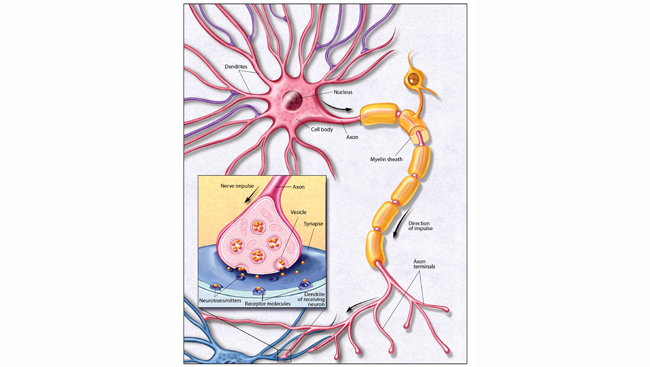

Post a Comment for "40 draw a labeled diagram of neuron"