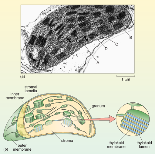
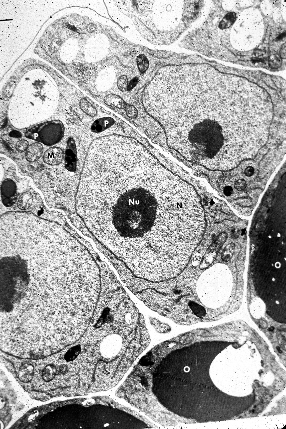
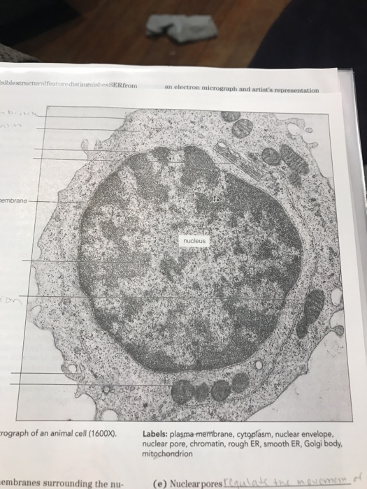
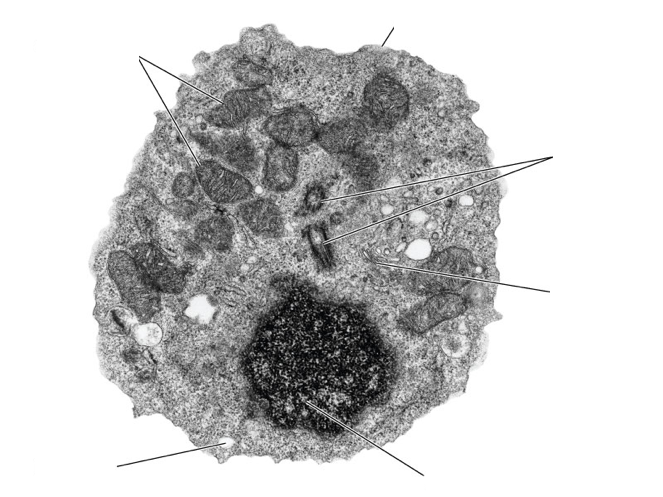
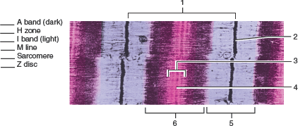
38 provide the labels for the electron micrograph
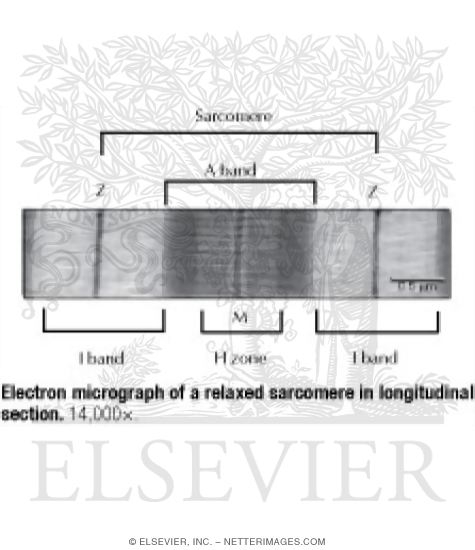
Electron Micrograph of a Relaxed Sarcomere In Longitudinal ... Please describe! how you will use this image and then you will be able to add this image to your shopping basket. Electron Micrograph of a Relaxed Sarcomere ... Electron micrograph Definition and Examples - Biology Online Dictionary It is not intended to provide medical, legal, or any other professional advice. Any information here should not be considered absolutely correct, complete, and up-to-date. ... Electron micrograph (Science: microscopy) a photographic reproduction of an image formed by the action of an electron beam. Electron microscope See: microscope, electron.
A Study of the Microscope and its Functions With a Labeled Diagram To better understand the structure and function of a microscope, we need to take a look at the labeled microscope diagrams of the compound and electron microscope. These diagrams clearly explain the functioning of the microscopes along with their respective parts. Man's curiosity has led to great inventions. The microscope is one of them.
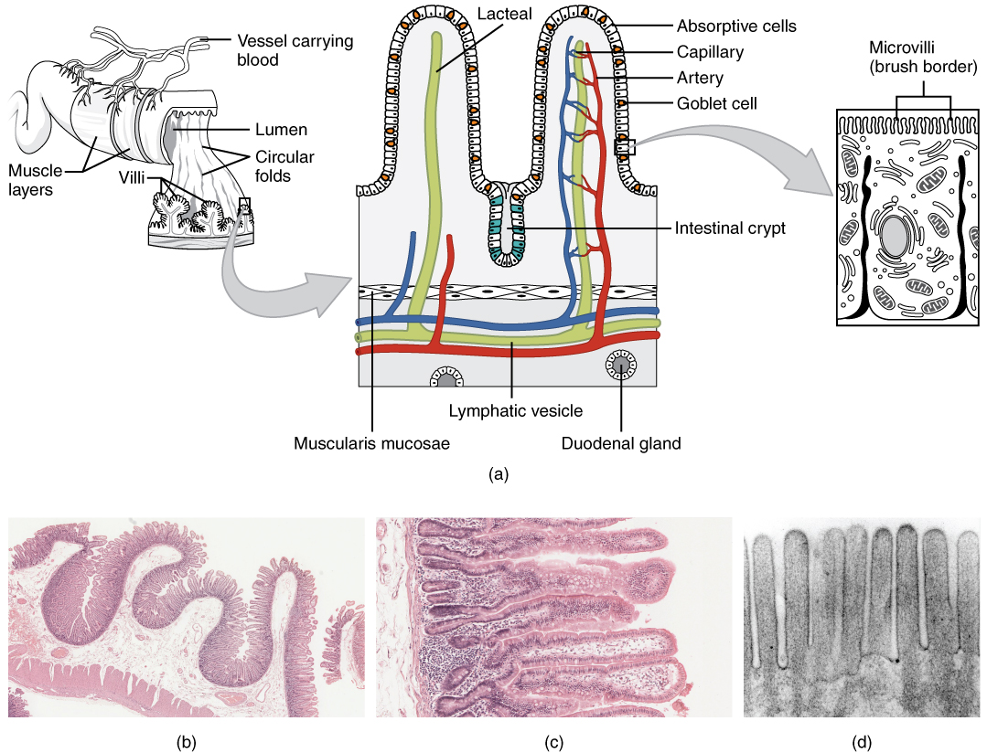
Provide the labels for the electron micrograph
Micrograph - an overview | ScienceDirect Topics The SEM micrograph of S41, shown in Fig. 5a, reveals the presence of regular spheres with diameters in the range between 500 - 1000 nm. For C41 (Fig. 5b) a sponge like structure with pores > 1 μm is observed. The micrograph of "as made" Z41(50) (Fig. 5c) shows roughly cubic crystals with rounded edges and a size in the range 500 - 800 nm. Provide the labels for the electron micrograph in figure 19.5. 8 ... Provide the labels for the electron micrograph in figure 19.5 Image transcription text 8 Human A&P | Lab Terms: A band (dark) emps I band (light) Sarcomere 2 Z line H zone M line 3 GURE 19.5 Using the terms provided, identify the bands and lines of the striations of a transmission electron crograph of relaxed sarcomeres (16,000x). Label This Transmission Electron Micrograph - Kaiden Brown Label This Transmission Electron Micrograph : TEM of chloroplast from Coleus blumei - Stock Image - B110 - Kaiden Brown To leave a comment, click the button below to sign in with Google. Sign in with Google Join Our Newsletter
Provide the labels for the electron micrograph. 16 The following electron micrograph shows part of a palisade mesophyll ... The electron micrograph shows a palisade mesophyll cell. a.i. State the name of the structures labelled I and II. [1] 1. Cell wall 2. Chloroplasts 3. Mitochondria. A. ii. Outline the function of the structure labelled III. [2] The mitochondria produce the energy necessary for the cell's survival and functioning. BSC2085L Lab 8 Exercises 12, 13, & 14 Flashcards | Quizlet Image: Provide the labels for the electron micrograph in figure 12.8. Label the following image using the terms provided. Terminal Cisternae, Thick and Thin ... Scanning electron microscope - Wikipedia A scanning electron microscope (SEM) is a type of electron microscope that produces images of a sample by scanning the surface with a focused beam of electrons.The electrons interact with atoms in the sample, producing various signals that contain information about the surface topography and composition of the sample. The electron beam is scanned in a raster scan pattern, and the position of ... Electron Micrographs of Cell Organelles | Zoology - Biology Discussion The Electron Micrograph of Lysosomes: This is the electron micrograph of Lysosome, and is characterized by following features. These are also called Suicide bags or Death bags of the cell (Fig. 13 &14): (1) They were discovered by de Duve (1954). (2) They are spherical or irregular membrane bound vesicles filled with digestive enzymes.
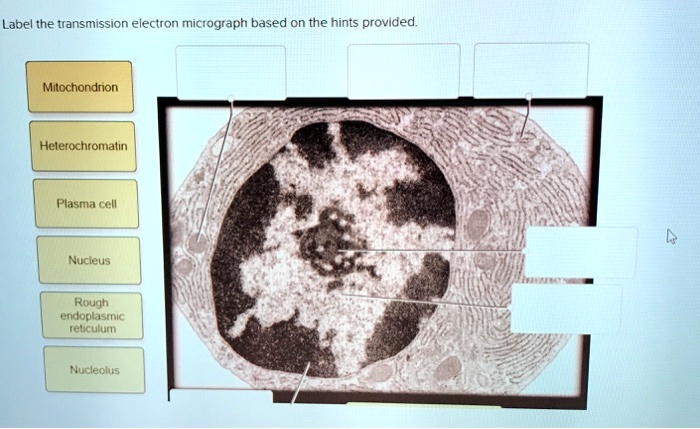
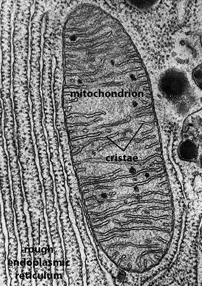
1.2 Skill: Interpretation of electron micrographs - YouTube Dec 25, 2015 ... Interpretation of electron micrographs to identify organelles and deduce the functions of specialized cells. Electron micrographs used with ... Electron Microscope- Definition, Principle, Types, Uses, Labeled Diagram Electron microscopes are used to investigate the ultrastructure of a wide range of biological and inorganic specimens including microorganisms, cells, large molecules, biopsy samples, metals, and crystals. Industrially, electron microscopes are often used for quality control and failure analysis. Virtual EM Micrograph List | histology - University of Michigan 021. Plasma Cell: This electron micrograph shows a typical secretory cell, a plasma cell, which secretes immunoglobulin protein. Many of the major types of cellular organelles are visible in this image. In the nucleus, areas of euchromatin and heterochromatin can easily be identified. Virtual Slide. Electron microscopes - Cell structure - Edexcel - BBC Bitesize The electron microscope. Electron microscopes use a beam of electrons instead of beams or rays of light. Living cells cannot be observed using an electron microscope because samples are placed in ...
Electron micrograph Definition & Meaning - Merriam-Webster The meaning of ELECTRON MICROGRAPH is a micrograph made with an electron microscope. What Is an Electron Microscope (EM) and How Does It Work? - VHA ... Today there are two major types of electron microscopes used in clinical and biomedical research settings: the transmission electron microscope (TEM) and the scanning electron microscope (SEM); sometimes the TEM and SEM are combined in one instrument, the scanning transmission electron microscope (STEM): Labels, Electron Microscopy Sciences | VWR Labels, Electron Microscopy Sciences | VWR Labels with expanded temperature range and freezable in liquid and vapor phase nitrogen. They adhere to most plastics, glass and metals without cracking, peeling or degrading.Labels identify, warn, organize, or provide instructions for items handled in any working environment. Chapter 20 Problem 1LAB Solution | Laboratory Manual For Human Anatomy ... Access Laboratory Manual for Human Anatomy & Physiology Main Version 3rd Edition Chapter 20 Problem 1LAB solution now. Our solutions are written by Chegg experts so you can be assured of the highest quality!
Electron Micrographs - University of Oklahoma Health Sciences Center Electron Micrographs Below is a collection of electron micrographs with labelled subcellular structures that you should be able to identify. Also, be sure to observe any electron micrographs which are made available in the laboratory by the instructor.
Chapter 20 Problem 1LAB Solution | Laboratory Manual For Human ... - Chegg Access Laboratory Manual for Human Anatomy & Physiology 2nd Edition Chapter 20 Problem 1LAB solution now. Our solutions are written by Chegg experts so you can be assured of the highest quality!
Label the microscope — Science Learning Hub Label the microscope Interactive Add to collection Use this interactive to identify and label the main parts of a microscope. Drag and drop the text labels onto the microscope diagram. eye piece lens diaphragm or iris coarse focus adjustment stage base fine focus adjustment light source high-power objective Download Exercise Tweet
Solved Label this transmission electron micrograph of | Chegg.com Question: Label this transmission electron micrograph of relaxed sarcomeres by clicking and dragging the labels to the correct location Sarcamere 1 band (light) ...
Electron Micrographs Figure 21 Epithelial cells often display extensive basal plasma membrane infoldings as observed in this electron micrograph: 1. Compartmentalized mitochondrion ...
13 Protein found within thick myofibril 14 A small bundle of muscle ... A small bundle of muscle fibersProvide the labels for the electron micrograph in ... in this transmission electron micrograph of relaxed sarcomeres(8,400x), ...
plant cell label electron micrograph Diagram | Quizlet plant cell label electron micrograph Diagram | Quizlet plant cell label electron micrograph 3.3 (3 reviews) + − Learn Test Match Created by June_bee743 Terms in this set (7) chloroplast ... cell wall ... plasma membrane ... golgi apparatus ... nucleus ... vacuole ... mitochondria ...
Electron microscope - Wikipedia An electron microscope is a microscope that uses a beam of accelerated electrons as a source of illumination. As the wavelength of an electron can be up to 100,000 times shorter than that of visible light photons, electron microscopes have a higher resolving power than light microscopes and can reveal the structure of smaller objects.
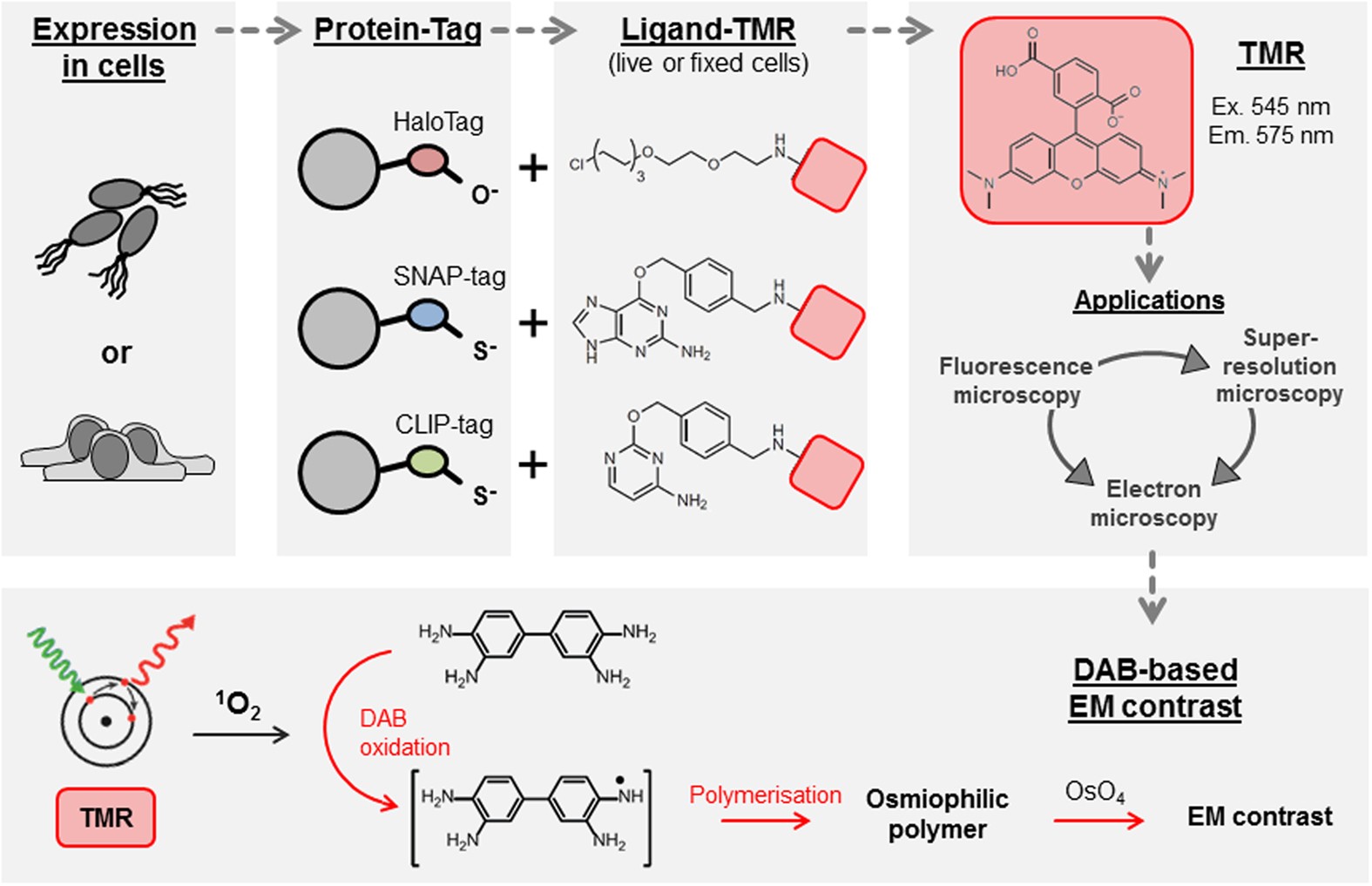
(A and B) Electron micrograph of a cell labeled for/5-tubulin followed... No microtubule labeling is evident. Bar, 2/zrn. from publication: Fluorescence photooxidation with eosin: a method for high resolution immunolocalization ...
Label This Transmission Electron Micrograph Of A Relaxed ... - Blogger Provide the labels for the electron micrograph in figure 18.5. (b) section through a muscle in the extended condition (140 % of whole muscle resting length). Label the following image using the terms provided. Label this transmission electron micrograph of relaxed sarcomeres by clicking and dragging the labels to the correct location .
Electron micrograph | definition of electron micrograph by Medical ... electron micrograph: [ mi´kro-graf ] 1. an instrument for recording very minute movements by making a greatly magnified photograph of the minute motions of a diaphragm. 2. a photograph of a minute object or specimen as seen through a microscope. electron micrograph a graphic reproduction of an object as viewed with an electron microscope.
SKELETAL MUSCLE STRUCTURE 11. Protein found within thick myofibril. 12. A small bundle of muscle fibers. PART B. Provide the labels for the electron micrograph in figure 19.4.
What is Electron Microscopy? - UMass Chan Medical School Electron microscopy is used in conjunction with a variety of ancillary techniques (e.g. thin sectioning, immuno-labeling, negative staining) to answer specific questions. EM images provide key information on the structural basis of cell function and of cell disease.
provide labels for the electron micrograph in albania Customized shelf label epaper,export e-paper electronic shelf label,top sale e-link electronic paper label,LCD price tags maker,electronic tag for price,electronic online labels tawian,high quality Supermarket smart price tag,e ink shelf label fast delivery,electronic shelf label samsung,electronic shelf label epop Exporters,export Magnetic Price Tag Holder,OEM led light demo kit,electrical ...
Diagram of labeling used for light and electron microscopy. A: Labeling ... An ideal label for light and electron microscopy would contain both fluorescent dye and a colloidal nanoparticle on a single antibody molecule. Secondary and tertiary label, the colloidal ...
Muscle Lab 19 Figure 19.5 Sarcomere Diagram - Quizlet Start studying Muscle Lab 19 Figure 19.5 Sarcomere. Learn vocabulary, terms, and more with flashcards, games, and other study tools.
RCPA - Electron microscopy Electron microscopy is useful for rapid viral identification and is important for typing Glomerulonephritis . In tumour diagnosis where results from morphology and antibody labelling are equivocal electron microscopy can provide useful additional diagnostic information. For the other conditions such as metabolic inherited disorders, peripheral ...
Solved Before Going to Lab Label the electron micrograph of - Chegg Transcribed image text: Before Going to Lab Label the electron micrograph of the sarcomere in Figure 12.5. nents in the etal muscle ghter color he I bands.
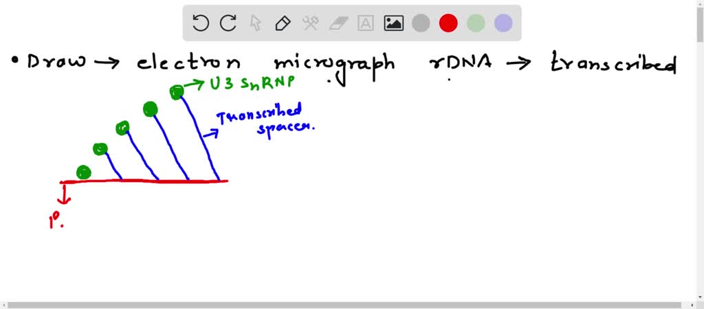
Draw a representation of an electron micrograph of rDNA being transcribed. Label the non-transcribed spacer, the transcribed spacer, the RNA polymerase molecules, the U3 snRNP, and the promoter.
Label This Transmission Electron Micrograph - Kaiden Brown Label This Transmission Electron Micrograph : TEM of chloroplast from Coleus blumei - Stock Image - B110 - Kaiden Brown To leave a comment, click the button below to sign in with Google. Sign in with Google Join Our Newsletter
Provide the labels for the electron micrograph in figure 19.5. 8 ... Provide the labels for the electron micrograph in figure 19.5 Image transcription text 8 Human A&P | Lab Terms: A band (dark) emps I band (light) Sarcomere 2 Z line H zone M line 3 GURE 19.5 Using the terms provided, identify the bands and lines of the striations of a transmission electron crograph of relaxed sarcomeres (16,000x).
Micrograph - an overview | ScienceDirect Topics The SEM micrograph of S41, shown in Fig. 5a, reveals the presence of regular spheres with diameters in the range between 500 - 1000 nm. For C41 (Fig. 5b) a sponge like structure with pores > 1 μm is observed. The micrograph of "as made" Z41(50) (Fig. 5c) shows roughly cubic crystals with rounded edges and a size in the range 500 - 800 nm.












Post a Comment for "38 provide the labels for the electron micrograph"